Madrid (EFE).- Twelve years is what scientists have taken to map the brain of an insect, first dividing it into 5,000 slices and then scrutinizing them and thus being able to reconstruct all its pieces, neuron by neuron. Why is it important to have the complete picture of a brain even if it is that of a small arthropod?
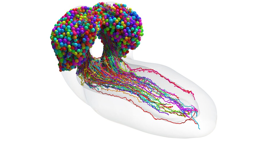
Until now, neuroscience has worked with partial maps or, in the best of cases, complete ones, but of species with a few hundred or thousands of neurons. The new neuronal atlas of the fruit fly larva, the most extensive to date, includes 3,016 neurons and all the connections between them, 548,000.
“Now we can start to study the brain as it is, its structure. We can, for example, try to understand how the integration of the senses works, where visual information comes together with olfactory and touch, or how memory is combined with new information that arrives”, explains Albert Cardona to EFE.
Cardona, from the MRC Laboratory of Molecular Biology in the United Kingdom and the University of Cambridge, is one of the researchers who on March 9 published in Science the complete brain connectome -diagram of neuronal connections- of the larva of “Drosophila melanogaster ” (vinegar or fruit fly).

meticulous method
The researchers, also from Johns Hopkins University, implemented a painstaking and collaborative method. It all began with the dissection of the larva to remove its nervous system with two tweezers and submerge it in a fixative liquid.
Then came the heavy metals, which were added to differentially stain cell membranes and proteins, and then converted to resin to be put in an oven to harden it and thus be able to use a machine capable of making very fine serial cuts -of 40 nanometers thick – with its diamond blade.
In total 5,000 slices, which were classified in order. “It is important not to mix them,” says the Spanish researcher.
One by one they were passed through the high-resolution electron microscope and then, thanks to software developed by Cardona, among others, the images were joined, in a continuous volume. Everything was uploaded to a server that the team accesses through a browser.
“Once there, the reconstruction work of all the neurons and connections began, cut by cut, to achieve the 3D image.”
The first attempt, in 1970
The first attempt to map a brain, a 14-year effort on the roundworm, began in the 1970s and resulted in a partial map and a Nobel Prize – for this and other achievements – for Sydney Brenner, John Sulston and Bob Horvitz.
The connectome now of the larva of the fly represents a very important step for neuroscience and will serve to understand the general architecture of the brain, not only that of this small animal, but also that of humans.
The fruit fly is also one of the leading models in science because it shares much of its fundamental biology with us. In this case, your brain map can help you understand neurodegenerative diseases.
For example, Parkinson’s appears when a type of neurons called dopaminergic neurons die and there is not enough amount of dopamine, a neurotransmitter that participates, among others, in motor behavior.
Mapping the brain of the fly larva induced with Parkinson’s and comparing it with one without the disease can help to understand what is happening in the brain circuits or why the tremors appear, and test drugs, Cardona stresses to EFE.
Benefits for AI
In addition to advancing knowledge of disease and human thought, the first insect brain atlas will inspire new machine learning architectures.
“We have found -on the map- things that we don’t know what they are for, but they are there and they surely have a function. The neural networks will end up copying it,” says Cardona, who received his doctorate from the University of Barcelona and has been in Cambridge for four years, after passing through Switzerland, California and Virginia (he began this work at the Howard Hughes Medical Institute and spent eight years).
Neurons have several branches, some are dendrites, which generally act as receivers of information, and other axons, which function as transmitters of information to other neurons.
Two thirds of the connections in the brain of the fly larva are like this, but in one third other schemes are repeated, such as axon-axon or dendrite-dendrite connections.
Although their existence is known, also in humans, the proportion is not known, nor is it known whether they are accidental or consistent and for what exact purpose.
There is still a lot to analyze and understand, Cardona stresses, and of course it is still necessary to make the leap to other animals, although the technology and the budget are still limited; it is estimated that scientists will not be able to deal with the mouse brain until the next decade.
However, he says, between larva and mouse there are very attractive things; His lab is mapping the brain of the dwarf squid.