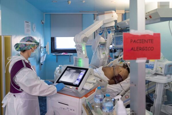
Barcelona (EFE).- The Hospital del Mar in Barcelona is the first center in Catalonia to implement a surgical program using sodium fluorescein, a dye that binds to cancer cells, to improve detection and resection of tumors in the brain.
The staining of cancer cells allows surgeons to obtain greater precision when performing complete resection of the tumor, in addition to guaranteeing that at the end of the surgery all traces of tumor have been eliminated, limiting the risk of recurrence and, therefore, , increase survival.
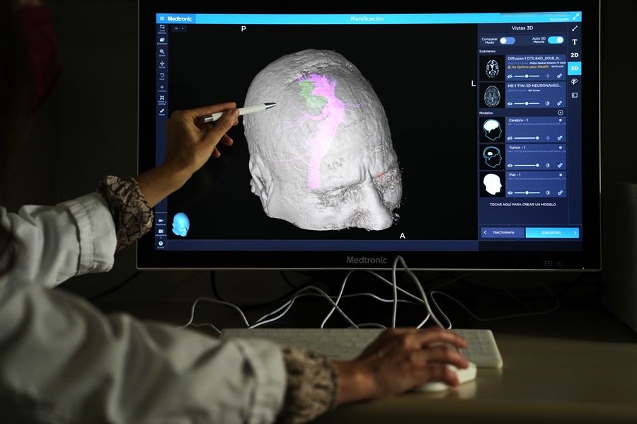
The neurosurgeon coordinator of the Hospital del Mar Neurosurgery Service and head of brain oncology surgery at the center, Gloria Villalba, explained that “fluorescence can provide very relevant information, as it can help us make malignant cells visible more beyond the tumor capsule, not otherwise visible.
“In addition, it also helps to confirm during the same surgery with the pathologist that they are tumor cells, and to remove the affected area, performing larger and more beneficial resections from the oncological point of view”, added Villalba.

According to the neurosurgeon, oncological guidelines indicate that it is necessary to leave a safety margin of healthy tissue beyond the capsule of the metastasis to guarantee that all malignant cells are removed, that is, that the resection should go between 4 and 5 millimeters beyond what is indicated by the images of the patient’s brain, although this standard does not guarantee its complete elimination.
With sodium fluorescein, which is injected by vein into the patient in the same operating room and has no side effects, it can be checked whether this margin is sufficient or whether it needs to be widened.
the tumor shines
Sodium fluorescein causes tumor cells to glow with yellow fluorescence if viewed through a microscope equipped with the appropriate filters and its usefulness has been proven in other pathologies, such as some ophthalmological ones, but until now it has been little used in neurosurgery. for brain metastases.
In other types of tumors, such as gliomas, there are other substances that fulfill this function, but they did not work in patients with metastases.

The Hospital del Mar team, which includes professionals from Neurosurgery, Pathology, Pharmacy, Medical Oncology, Radiation Oncology, Neurology and Radiology, began working with this dye at the end of 2022.
In the operating room, the surgeons take samples from the area illuminated by the sodium fluorescein and send them to the Pathology Service to certify that they are tumor cells and to continue resection of the tumor.
“This means that the probability of recurrence is much lower, since it depends on the tumor cells that may have remained after surgery,” highlighted Villalba and Alejandra Narváez, an assistant physician at the Neurosurgery Service, both in charge of brain metastasis in the center.
In this sense, doctors Montserrat Arumí and Gina Parini, associate physicians of the Pathological Anatomy Service, have indicated that they make intraoperative consultations while surgery is carried out with sodium fluorescein.
“Samples are sent to us from the operating room, during the surgical act, which are quickly processed using frozen sections to be able to make a diagnostic orientation and thus be able to decide the most appropriate therapeutic option”, they have reiterated.
In this way, “in the cases in which sodium fluorescein is used, we have observed, in samples from tissue adjacent to the lesion, in apparently unaffected areas, the presence of few groups of tumor cells infiltrating the tissue. Given the results observed, we believe that it can be very useful to be able to perform a more complete excision of the tumor”, they have concluded.