Pamplona (EFE).- The Vascular Surgery Service of the University Hospital of Navarra (HUN) has become the first in Spain to carry out a complex procedure to solve a lesion that a patient had in the aorta artery.
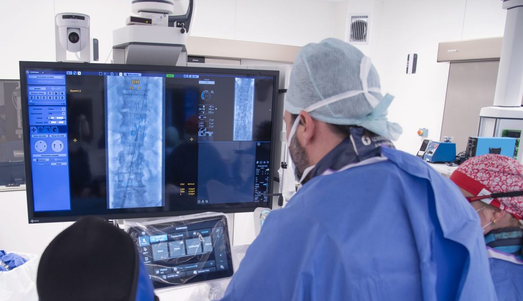
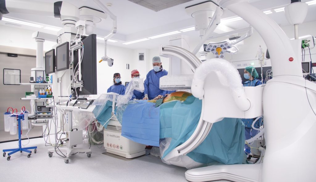
The procedure, known as endovascular aortic repair using a transcaval approach, took place on February 27 and consisted of placing a prosthesis in the aorta, navigating with a series of guidewires and catheters through an adjacent vein, the vena cava, and penetrating into the aorta after making an access through the walls of both blood vessels.
As detailed by the Provincial Government, despite the complexity of the intervention and its apparent aggressiveness, it is a minimally invasive technique, which has favored the recovery of the patient, who remains hospitalized, fully recovered from surgery and waiting to be given discharged in a few days to continue monitoring on an outpatient basis.
An unprecedented technique in Spain
The procedure was intended to cover an ulcer located in the thoracic aorta, the upper segment of this artery, the main one in the human body, which descends from the heart to the abdomen, where, at the level of the pelvis, it divides into two, the iliac arteries.
The usual practice in this type of intervention, as indicated, consists of introducing a catheter with the prosthesis, accessing one of these arteries at the level of the groin and going up that vessel until reaching the affected area.
In this case, however, the patient had an occlusion both of both iliac arteries and above, at the level of the infrarenal portion of the aorta, which made access by the ordinary route impossible.
In this situation, the team of vascular surgeons, led by doctors Sebastián Fernández and Esther Martínez, considered the possibility of transcaval access, reported in the medical literature, but for which there were no precedents in Spain for a repair of these characteristics.
The vena cava is anatomically located next to the aorta throughout its entire length, both in the thorax and in the abdomen, but they are not communicated, since they are different blood circuits.
The surgeons entered the femoral vein, also at the level of the groin, and ascended until they reached the cava, to then cross its wall and that of the aorta. Once inside this artery, they continued to raise the device up to the affected area, in order to implant the endoprosthesis and “reinforce” the area of the ulcer.
Likewise, to control the procedure, the artery of the right arm was approached. Once the endoprosthesis was placed, the communication hole made in the vessels was closed using a self-expanding vascular plug.
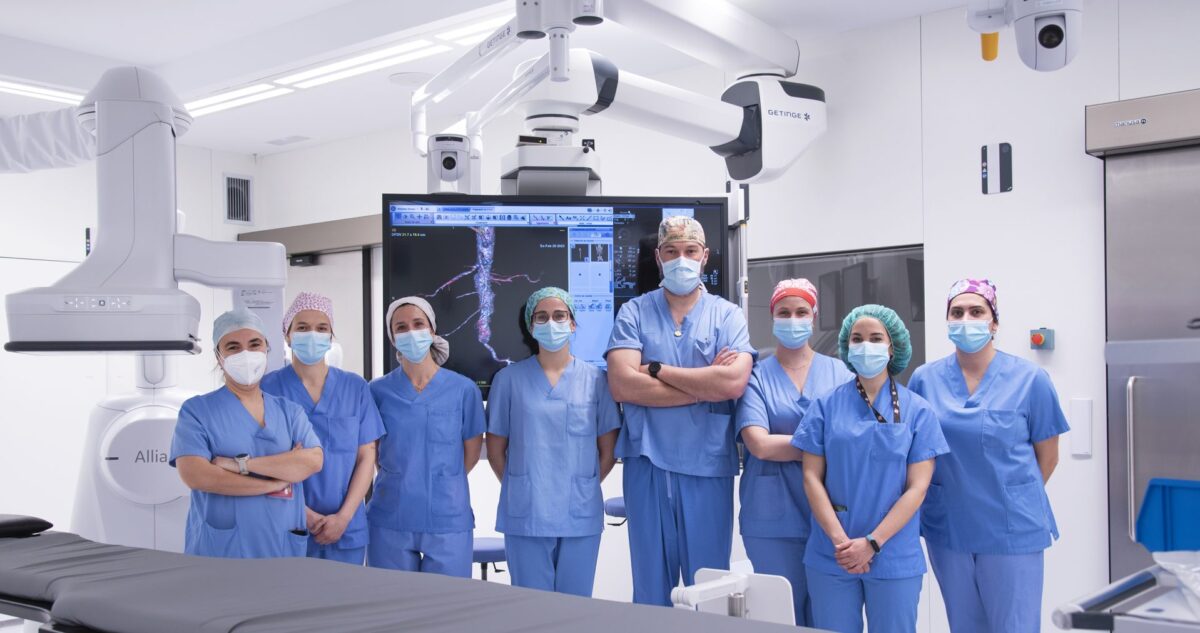
Both in the diagnosis and in the post-surgery follow-up, the Internal Medicine Service has also participated and, especially, Dr. María Gracia Ruiz de Alda.
In addition to doctor Gracia, and doctors Sebastián Fernández and Esther Martínez, nurses Meritxell González, María Esquíroz and Lucía Ursúa have intervened; the specialist physician in Vascular Surgery, Amaya Arróniz; the anesthetist, Orreaga Zugasti; the techniques of Rayos, Andrea Monreal and Nieves Martínez; and the medical specialists in Anesthesiology and Resuscitation, Alejandro Bilbao and Andrés Alegre.
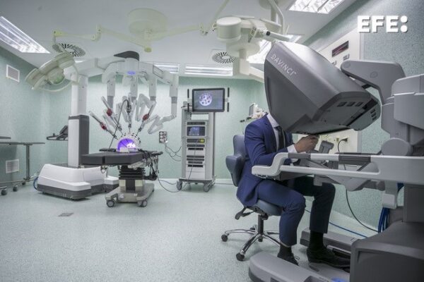
The Provincial Government highlights that the performance of this technique has benefited to a great extent from the technological advances offered by the new hybrid operating room for vascular surgery that came into operation at the HUN last May.